
Brain AVM (Arteriovenous Malformation) — What You Need to Know
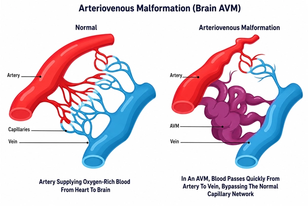
A brain arteriovenous malformation, or AVM, is an abnormal tangle of blood vessels in the brain. It is usually present from birth. In a healthy brain, arteries deliver blood at high pressure to a network of tiny vessels called capillaries, which slow the flow before it passes into the veins. In an AVM, this normal step is missing: arteries connect directly to veins through a tangle of vessels, so blood rushes through at high pressure without being slowed down.
How a brain AVM differs from normal blood vessels
The key difference is the missing capillary network. Because the small vessels that normally cushion the flow are absent, the veins in an AVM are exposed to a much higher pressure than they are built to handle. Over time this can stretch and weaken the vessels, which is what makes an AVM a concern. The central tangle of abnormal vessels is often called the nidus.
Are brain AVMs present from birth?
In most cases an AVM is congenital, meaning it is present from birth, although it may not be discovered until much later in life — or sometimes not at all. AVMs are uncommon, and many people who have one never develop symptoms. When symptoms do appear, they often show up in early or middle adulthood.
Symptoms and warning signs
Often no symptoms until discovered
Many AVMs cause no symptoms at all and are found by chance during a scan carried out for an unrelated reason. When an AVM is discovered this way, it can be assessed carefully to understand its size, location, and pattern of blood flow.
Seizures
Seizures are one of the more common ways an AVM first makes itself known. They occur because the abnormal vessels can irritate the surrounding brain tissue.
Headaches
Some people with an AVM experience headaches. These can vary widely from person to person and are not specific to AVMs, but a persistent or unusual headache is always worth discussing with a doctor.
Bleeding (hemorrhage) — the most serious risk
The most serious concern with an AVM is that the weakened vessels may rupture and bleed into or around the brain. This is a medical emergency. It can cause a sudden severe headache, weakness, difficulty speaking, or loss of consciousness, and requires emergency care immediately.
Neurological symptoms depending on location
Because different parts of the brain control different functions, an AVM can sometimes cause focal symptoms based on where it sits — for example, weakness, numbness, vision changes, or problems with balance or coordination.
How a brain AVM is diagnosed
An AVM is diagnosed using imaging that shows the brain’s blood vessels and the pattern of blood flow through them. Often more than one type of scan is used together to build a complete picture.
MRI and MR angiography
MRI shows the brain tissue in detail and can reveal an AVM and any effect it is having on the surrounding brain. MR angiography maps the blood vessels themselves, helping to show the size and position of the malformation.
CT and CT angiography
A CT scan is quick and is especially useful in an emergency to check for bleeding. CT angiography adds a detailed map of the blood vessels to help locate the AVM.
Digital subtraction angiography (DSA)
DSA is considered the most detailed test for an AVM. A thin catheter is guided to the brain’s arteries and contrast dye is injected while X-ray images are taken. It shows the flow of blood through the AVM moment by moment, which is important for understanding its structure and planning any treatment.
How brain AVMs are assessed and graded
To help understand an AVM and weigh up treatment options, doctors often use a grading system — most commonly the Spetzler-Martin scale. It considers three features of the AVM to give an overall picture of how complex it is. This is used by the medical team as a planning tool and is explained here only for general understanding.
- Size of the AVM — how large the tangle of vessels is.
- Location — whether it sits in or near a part of the brain that controls important functions such as movement, speech, or vision.
- Venous drainage — the pattern of the veins carrying blood away from the AVM.
Together these factors help the team understand the balance of risks and benefits for each possible approach, which differs from person to person.
Treatment approaches for brain AVMs
There is no single treatment that suits every AVM. The right approach — or the decision to simply monitor — depends on the AVM’s size, location, and blood flow pattern, whether it has bled, the symptoms it is causing, and the person’s overall health. Sometimes more than one method is combined. The options below are explained for understanding only; any decision is made together with the treating team.
Observation and monitoring
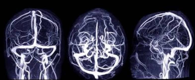
For some AVMs, particularly those that are not causing symptoms and sit in high-risk locations, careful monitoring over time may be the safest course. This means keeping the situation under review rather than intervening straight away.
Endovascular embolization — how it works
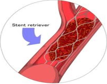
This is a minimally invasive approach carried out from inside the blood vessels. A thin catheter is guided through the arteries to the AVM, and a special material is used to block off the abnormal vessels, reducing the flow of blood through the malformation. It is sometimes used on its own and sometimes as a preparatory step before surgery or radiosurgery.
Stereotactic radiosurgery — how it works
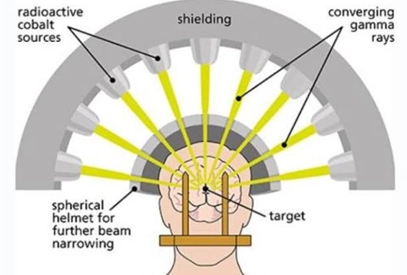
Despite its name, radiosurgery does not involve an incision. It uses precisely focused beams of radiation directed at the AVM to make its vessels gradually close off over a period of months to a couple of years. It may be considered for smaller AVMs or those in locations that are difficult to reach.
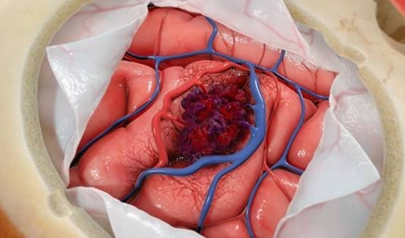
Microsurgical removal — when it may be considered
This is an open surgical operation to remove the AVM directly. It may be considered for AVMs that are accessible and where the balance of risks favours removal. It is one of several approaches a team may weigh up for a given case.
When to seek immediate medical attention
Some signs may indicate that an AVM has bled, which is a medical emergency. Seek emergency care without delay if there is:
- A sudden, severe headache unlike any experienced before
- A seizure, especially a first-ever seizure
- Sudden weakness, numbness, or difficulty speaking
- Sudden vision changes or loss of balance
- Loss of consciousness, even briefly
These symptoms need emergency assessment immediately. For an AVM found by chance, or for ongoing concerns, speak to a specialist.
Frequently Asked Question
Is a brain AVM the same as a brain aneurysm?
No. An aneurysm is a bulge in the wall of a single blood vessel, while an AVM is an abnormal tangle where arteries connect directly to veins without the normal small vessels in between. They are different conditions, although both involve the brain’s blood vessels and both are assessed with detailed imaging.
Can you be born with an AVM and never know it?
Yes. Most AVMs are present from birth, and many never cause symptoms. Some are found only by chance on a scan done for another reason, and a number are never discovered at all. When symptoms do appear, they often show up in early or middle adulthood.
What is the main risk of a brain AVM?
The most serious risk is that the weakened vessels rupture and bleed into or around the brain, which is a medical emergency. Other ways an AVM can show itself include seizures and headaches. The level of risk varies from person to person.
Does every AVM need to be treated?
Not always. For some AVMs, particularly those causing no symptoms or sitting in high-risk locations, careful monitoring may be the safest option. Treatment decisions weigh the AVM’s features against the risks and benefits of each approach, and are made by the treating team with the patient.
What does AVM grading mean?
Grading, such as the Spetzler-Martin scale, is a way of describing how complex an AVM is by looking at its size, location, and venous drainage. It is a planning tool used by the medical team to understand the balance of risks and benefits, and is explained here only for general understanding.