
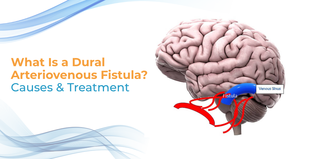
A dural arteriovenous fistula and a brain AVM are not the same condition. A dAVF is a faulty link between an artery and a vein in the membranes covering the brain, and it usually develops later in life. An AVM sits within brain tissue and is present from birth. The distinction matters because the drainage pattern, not the size or location alone, determines how dangerous a fistula is and how it must be treated.
According to Dr. Savyasachi Jain, an Interventional Neuroradiology specialist, “A dAVF is graded by where its blood drains, not by how big it is. When flow refluxes backward into the cortical veins, the risk of bleeding rises sharply. That single finding decides whether a fistula is watched or treated.”
What causes a dAVF and what symptoms does it cause?
Most dAVFs are not present at birth. They appear later in life. A blocked or narrowed venous sinus is the usual starting point, and pressure builds behind it until abnormal links open between nearby arteries and the vein.
- Triggers: A head injury. Past surgery. An infection, or a clot in a sinus. Often no cause is found at all.
- Pulsatile tinnitus: A rhythmic whoosh in one ear, timed to the pulse. Frequently the only clue.
- Headache and eye changes: New or persistent headache. A red, bulging, or pulsing eye points toward the cavernous sinus.
- Neurological signs: Reflux into brain veins is the worrying pattern. Seizures, weakness, or a bleed may follow.
These early signs can mimic a brain stroke. Sudden new symptoms are treated as urgent from the start.
How is a dAVF diagnosed and treated?
Imaging confirms the fistula and maps the drainage. That drainage pattern decides everything that follows.
- Imaging: MRI and MR angiography usually come first. CT angiography helps in emergencies. DSA settles the diagnosis and shows the grade.
- Observation: A low-grade fistula without cortical reflux may simply be watched. This suits cases where tinnitus is the only complaint.
- Endovascular embolization: The main approach. A catheter reaches the fistula from inside the vessels and seals the faulty link.
- Surgery and radiosurgery: Surgical disconnection is kept for fistulas other methods cannot reach. Radiosurgery closes selected ones slowly.
Many high-grade fistulas can be cured outright. The same watch-or-treat judgement applies to a brain aneurysm, weighed one case at a time.
A dAVF is graded by its drainage, not its symptoms. Sudden severe headache, seizure, or new weakness needs emergency care.
Why Choose Dr. Savyasachi Jain?
Dr. Savyasachi Jain is an Interventional Neuroradiologist and Assistant Professor in Neuroimaging and Interventional Neuroradiology at AIIMS, New Delhi. His work covers vascular conditions of the brain, spine, and head and neck, dural arteriovenous fistulas among them, using image-guided endovascular techniques.
A dAVF has to be graded accurately before any treatment decision is made. That grading rests on detailed vessel imaging and the experience to read it correctly. This page is offered as an educational resource on the condition and is not advice for any individual case.
Frequently Asked Questions
Is a dAVF the same as a brain AVM?
No. A dAVF forms in the brain’s coverings and usually develops later in life. An AVM is a tangle within brain tissue, typically present from birth.
Is pulsatile tinnitus always caused by a dAVF?
No. It has several causes. But a heartbeat-like sound in one ear should be assessed, since a dAVF is one treatable possibility.
Are all dAVFs dangerous?
No. Low-grade fistulas without reflux into brain veins are often benign. Risk rises when drainage backs up into cortical veins.
Can a dAVF be cured?
Yes. Many are closed completely, most often through endovascular embolization.
References
Disclaimer
This blog is for educational and informational purposes only and should not be considered professional advice.